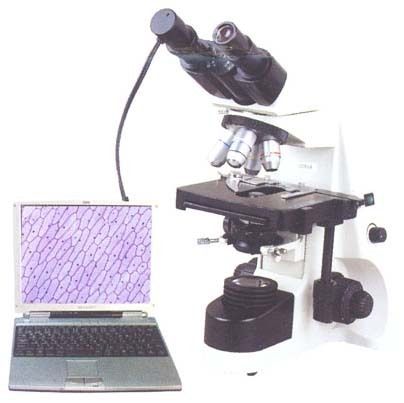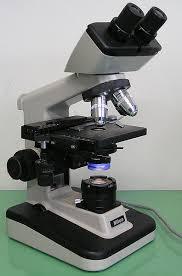


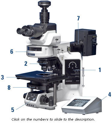
Microscopes - Electron And Optical Innovations | Advanced Imaging, High Resolution, Superior Magnification
Get Latest Price

In Stock
Product Specifications
| Magnification | 1000x+ |
| Resolution | Sub-micron |
| Type | Optical |
| Power | 100-240V |
| Weight | 20kg+ |
| Dimensions | 50x40x60cm |
| Usage | Microscopic cell analysis, sample examination, detailed observation and measurement of specimens |
| Material | Metal,Glass |
| Features | High resolution, Precise imaging, Detailed analysis, Efficient workflow |
Product Overview
Key Features
It was not until the 1660s and 1670s that the microscope was used extensively for research in Italy, The Netherlands and England. Marcelo Malpighi in Italy began the analysis of biological structures beginning with the lungs. Robert Hooke'sMicrographia had a huge impact, largely because of its impressive illustrations. The greatest contribution came fromAntonie van Leeuwenhoek who discovered red blood cells and spermatozoa and helped popularise microscopy as a technique. On 9 October 1676, Van Leeuwenhoek reported the discovery of micro-organisms
Electron microscopy[edit]
An ant as imaged using a scanning electron microscope (SEM)
See also: electron microscope
In the early 1900s a significant alternative to light microscopy was developed, using electrons rather than light to generate the image.Ernst Ruska started development of the first electron microscope in 1931 which was the transmission electron microscope (TEM). The transmission electron microscope works on the same principle as an optical microscope but uses electrons in the place of light and electromagnets in the place of glass lenses. Use of electrons instead of light allows a much higher resolution.
Development of the transmission electron microscope was quickly followed in 1935 by the development of the scanning.
Company Details
Focusing on a customer-centric approach, PRAXOR INSTRUMENTS & SCIENTIFIC CO has a pan-India presence and caters to a huge consumer base throughout the country. Buy Microscope in bulk from PRAXOR INSTRUMENTS & SCIENTIFIC CO at Trade India quality-assured products.
Business Type
Exporter, Manufacturer, Distributor, Supplier, Wholesaler
Employee Count
12
Establishment
2012
Working Days
Monday To Sunday
GST NO
33BPRPP0829K1ZG
Payment Mode
Cash on Delivery (COD), Cash Advance (CA)
Related Products
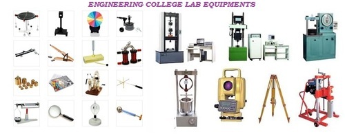
Educational Lab Equipment - General Use: Laboratory
Price: 5000 INR
MOQ - 1 Piece/Pieces
Subitek
Coimbatore, Tamil Nadu
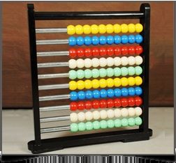
Educational Wooden Abacus Toy - Wood & Plastic, 10x15x2 inches, Black | 100 Beads, Early Learning, Easy Counting
Bharat Flow Controls
Coimbatore, Tamil Nadu
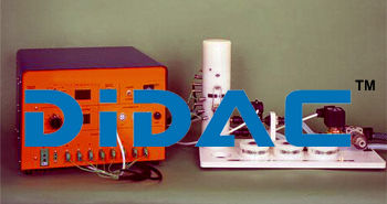
Computer Controlled Heat Transfer Teaching Equipment - Application: Laboratory
MOQ - 1 Pack/Packs,
Didac International
New Delhi, Delhi
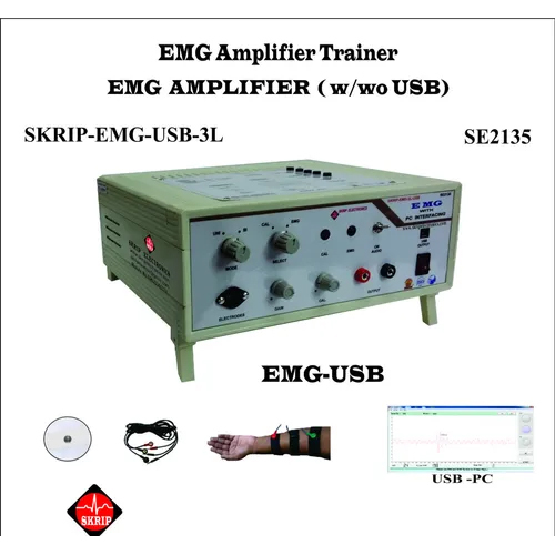
Emg Trainer (Electromyography) Usb Educational Hospital Study Application: Industrial
Price: 32000 INR
MOQ - 1 Unit/Units
Skrip Electronics
Mumbai, Maharashtra
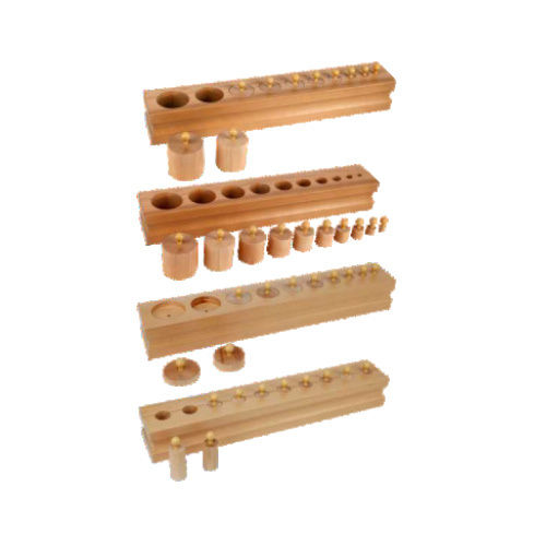

Outdoor Science Park Equipment - Material: Iron
Price: 17500.0 INR
MOQ - 1 Piece/Pieces
Wissen Techus Private Limited
Delhi, Delhi
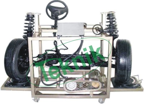
Independent Suspension Educational Equipment - Color: Black
MOQ - 1 Piece/Pieces
Micro Teknik
Ambala Cantt, Haryana
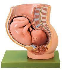

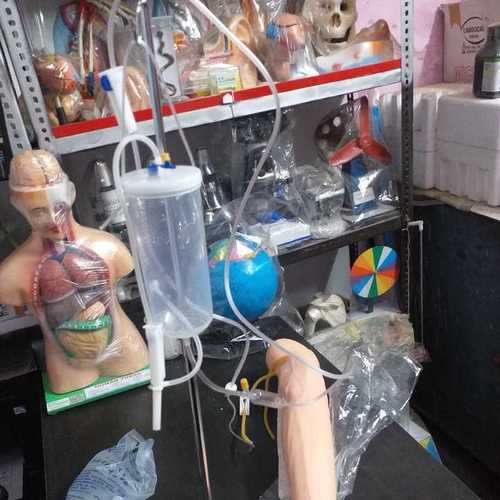
LABCARE EDUCATIONAL EQUIPMENT IN DEHRADUN
Price: 100 INR
Labcare Instruments & International Services
Ambala Cantt, Haryana
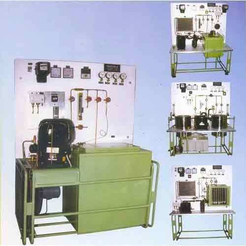
Mild Steel Educational Training Tools & Equipment
Price: 30000 INR
MOQ - 1 Unit/Units
Frigtools Refrigeration And Engineering Co.
Delhi, Delhi
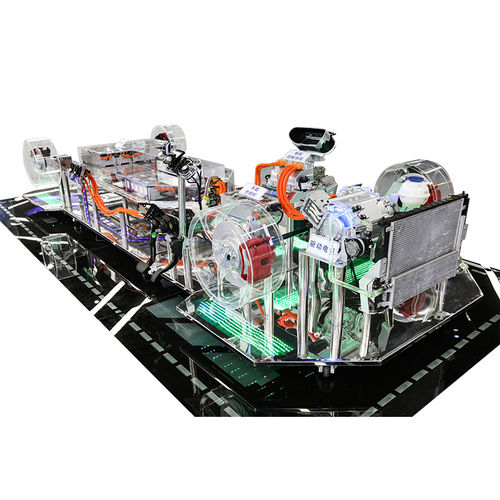
Grey Transparent Car Chassis Vocational Education Equipment
Beijing Zhiyang Beifang International Education Technology Co., Ltd
Beijing, Beijing
Explore Related Categories


More Products From This Seller






Seller Details


GST - 33BPRPP0829K1ZG
Chennai, Tamil Nadu
Partner
Mr Praveen Kumar Prakash
Address
141, Metro Nagar 3rd Avenue, Alapakkam, Porur, Chennai, Tamil Nadu, 600116, India
educational equipment in Chennai
Report incorrect details